


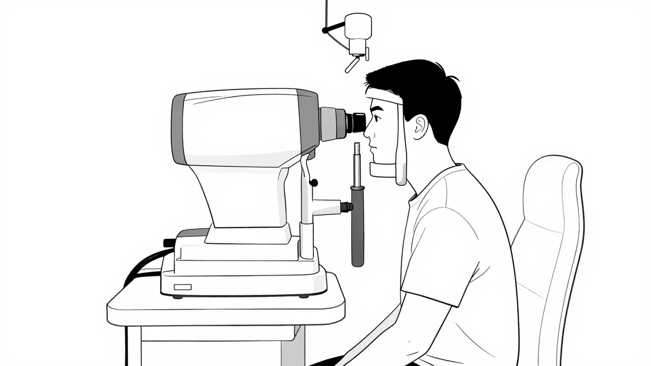
Imagine being able to see the intricate wiring of your brain without opening your skull. Now imagine doing that for your eyes. This is exactly what modern ophthalmic imaging allows doctors to do. If you have ever sat in a dark room while a machine clicked and whirred near your face, you were likely undergoing one of three critical tests: Optical Coherence Tomography (OCT), fundus photography, or angiography.
These are not just pictures; they are diagnostic powerhouses. They reveal hidden leaks, tiny blockages, and structural changes long before you notice any vision loss. Understanding the difference between these technologies helps you navigate your eye health with confidence. You will know why your doctor orders specific scans and what those images actually mean for your treatment plan.
The Snapshot: Fundus Photography
Think of fundus photography as the standard snapshot of your eye’s interior. It captures a 2D image of the back of the eye, known as the posterior pole. This area includes the retina, optic disc, macula, and blood vessels.
Specialized cameras, such as the Zeiss FF 450+, take these high-resolution photos. The process is quick, non-invasive, and requires no drops or injections. Doctors use these images primarily for documentation and baseline comparison. If you have diabetes or high blood pressure, these photos track changes over time.
However, a photo has limits. It shows surface details but cannot see inside the layers of the retina. It is like looking at a house from the street-you can see the paint and windows, but you cannot tell if the plumbing is leaking behind the walls. For deeper insights, we need more advanced tools.
The Cross-Section: Optical Coherence Tomography (OCT)
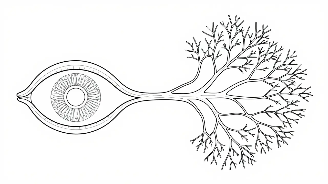
Optical Coherence Tomography, commonly called OCT, changes the game by providing cross-sectional views of the retina. Instead of a flat picture, OCT creates a slice-like image, similar to an ultrasound but using light instead of sound waves.
This technology allows doctors to visualize the distinct layers of the retina, including the retinal pigment epithelium, choroid, and vitreous. Spectral-domain OCT (SD-OCT) became the industry standard in the late 2000s because it offers incredibly high resolution-between 5 and 7 micrometers axially. To put that in perspective, this is detailed enough to see individual cells.
Why does this matter? Conditions like macular holes, diabetic macular edema, and glaucoma affect the structure of these layers. SD-OCT detects fluid accumulation or thinning that fundus photos miss entirely. More recently, swept-source OCT (SS-OCT) has emerged. SS-OCT performs 100,000 to 400,000 A-scans per second, compared to SD-OCT’s 20,000 to 85,000. This speed allows for deeper penetration into the choroid and faster acquisition, reducing motion blur.
The Blood Flow Map: Fluorescein Angiography (FA)
While OCT shows structure, fluorescein angiography (FA) reveals function-specifically, how blood flows through your retinal vessels.
Developed in the 1960s, FA remains the gold standard for detecting leakage. During the procedure, a nurse injects fluorescein dye into a vein in your arm. As the dye travels to your eyes, a specialized camera takes rapid photos. The dye lights up the blood vessels, allowing doctors to spot leaks, blockages, or abnormal new vessel growth.
This test is crucial for diagnosing wet age-related macular degeneration (AMD) and proliferative diabetic retinopathy. However, it is invasive. Some patients experience nausea, skin discoloration, or rare allergic reactions. The process also takes 10 to 30 minutes, requiring you to sit still while the dye circulates. Despite these drawbacks, FA provides unique information about leakage patterns that other methods cannot replicate.
The New Contender: OCT Angiography (OCTA)
OCT angiography (OCTA) represents a significant leap forward. Commercial systems arrived around 2014-2015, offering a non-invasive alternative to dye-based angiography.
OCTA uses the same hardware as OCT but employs sophisticated algorithms to detect the movement of red blood cells. It generates 3D maps of the retinal and choroidal vasculature without any injection. The scan takes only seconds. This speed and safety profile make it highly attractive for patients and clinicians alike.
OCTA visualizes superficial, middle, and deep capillary plexuses separately. It can identify enlarged foveal avascular zones (FAZ), microaneurysms, and neovascularization elsewhere (NVE). In studies comparing SD-OCTA and SS-OCTA, researchers found high correlation (r=0.86) in detecting abnormalities, though values are not interchangeable due to systematic differences in scanning speeds and depths.
Comparing the Technologies: Strengths and Weaknesses
No single imaging modality is perfect. Each has specific strengths and limitations depending on the condition being diagnosed. Here is how they stack up in clinical practice:
| Feature | Fundus Photography | Fluorescein Angiography (FA) | OCT / OCTA |
|---|---|---|---|
| Invasiveness | Non-invasive | Invasive (IV Dye) | Non-invasive |
| Primary Use | Documentation & Surface View | Detecting Leakage & Perfusion | Anatomical Layers & Vascular Flow |
| Acquisition Time | Seconds | 10-30 Minutes | Seconds |
| Sensitivity to Edema | Low | High (Gold Standard) | High (for fluid volume) |
| Limitations | No depth info | Allergic risk, time-consuming | Motion artifacts, misses subtle leakage |
In diabetic macular edema (DME), for example, FA demonstrates higher sensitivity (1.00) than SD-OCT (0.79) for detecting mild leakage. Small leaks may not create visible fluid pockets on OCT, leading to an 18% discrepancy rate in some studies. Conversely, OCTA excels in identifying non-perfusion areas in conditions like punctate inner choroidopathy (PIC), where traditional imaging often fails to show choriocapillaris defects.
Clinical Scenarios: Which Test Do You Need?
Your doctor chooses the imaging tool based on your symptoms and medical history. Let’s look at common scenarios.
Diabetic Retinopathy: If you have diabetes, regular monitoring is essential. Fundus photos screen for early signs. If abnormalities appear, OCT measures swelling (edema) thickness to guide treatment. OCTA helps detect neovessels (new, fragile blood vessels) without dye, which is safer for frequent monitoring. However, if severe leakage is suspected, FA may still be required to map the exact extent of damage.
Age-Related Macular Degeneration (AMD): For dry AMD, OCT tracks drusen deposits and retinal thinning. For wet AMD, where abnormal vessels grow under the retina, OCTA identifies the location and size of these vessels quickly. FA confirms active leakage, which determines whether anti-VEGF injections are needed.
Coats Disease: This rare condition involves abnormal blood vessel development. OCT reveals exudates in multiple retinal layers and subretinal fluid pockets that photos miss. Studies show OCT detects hyperreflective dots corresponding to macrophages and cholesterol crystals, linking imaging findings directly to histopathology.
Practical Considerations for Patients
Understanding the workflow helps reduce anxiety. Fundus photography and OCT/OCTA are quick and painless. You simply rest your chin on a stand and look at a target light. The main challenge with OCTA is staying still. Motion artifacts can ruin the image, making interpretation difficult. Children or patients with poor fixation may struggle with this.
FA requires an IV line. Afterward, your urine may turn yellow-orange for a day as the dye exits your body. Rarely, patients experience hives or breathing difficulties. Inform your doctor if you have allergies or kidney issues, as dye clearance depends on renal function.
Interpretation expertise matters too. While OCT reading is now standard in training, OCTA requires additional skill to distinguish true vascular anomalies from shadows or artifacts. Always ensure your specialist is experienced with the specific technology used.
Future Directions in Eye Imaging
Technology continues to evolve. Swept-source OCT improves choroidal visualization, aiding in the diagnosis of inflammatory conditions. Artificial intelligence (AI) algorithms are being integrated to automate the detection of diabetic retinopathy and glaucoma, potentially screening millions of images rapidly. Wide-field OCTA expands the view beyond the central retina, catching peripheral problems earlier.
Despite these advances, multimodal imaging remains the standard. Combining fundus photos, OCT, FA, and OCTA provides a comprehensive view of eye health. No single tool replaces the others yet. Instead, they work together to give your doctor the clearest possible picture of what is happening inside your eyes.
Is OCT painful?
No, OCT is completely painless and non-invasive. It uses light waves to create images, so there is no contact with your eye. You may feel a slight air puff if dilation drops are used beforehand, but the scan itself is gentle.
What is the difference between OCT and OCTA?
OCT shows the anatomical structure of the retina in cross-section, revealing layers and fluid. OCTA adds a layer of analysis to visualize blood flow within those layers without dye. Think of OCT as seeing the pipes, and OCTA as seeing the water moving through them.
Do I need dye for all eye scans?
No. Only fluorescein angiography (FA) requires an intravenous dye injection. Fundus photography, OCT, and OCTA are all non-invasive and do not involve needles or contrast agents.
How long does a fluorescein angiography take?
The entire process typically takes 10 to 30 minutes. This includes inserting the IV, injecting the dye, and capturing the series of photos as the dye circulates through your retinal vessels.
Can OCT detect early glaucoma?
Yes, OCT is highly effective at detecting early glaucoma. It measures the thickness of the retinal nerve fiber layer (RNFL) and the optic nerve head. Thinning in these areas often occurs before vision loss becomes noticeable to the patient.
Why might my doctor order both OCT and FA?
They provide complementary information. OCT shows structural damage and fluid volume, while FA identifies active leakage sources. For conditions like wet AMD or diabetic macular edema, knowing both the structure and the leak location helps tailor treatment precisely.
Are there risks associated with OCTA?
OCTA is safe and non-invasive. The main limitation is motion artifact. If you move during the scan, the image quality degrades. There are no radiation or chemical risks involved.
What is swept-source OCT (SS-OCT)?
SS-OCT is an advanced version of spectral-domain OCT. It scans faster (up to 400,000 A-scans per second) and penetrates deeper into the eye. This allows for better visualization of the choroid and reduces motion blur, making it ideal for complex cases.